In my last post I summarized the main routes by which different forms of actual or apparent motion can influence fMRI data. In the next few posts, I want to dig a little deeper into non-neural causes of variation in fMRI data. I am particularly interested in capturing information on the state of the subject at the time of the fMRI experiment. What else can be measured, and why might we consider measuring it? Brains don't float in free space. They have these clever life support systems called bodies. While most neuroimagers reluctantly accept that these body things are useful for providing glucose and oxygen to the brain via the blood, bodies can also produce misleading signatures in fMRI data. My objective in this series of posts is to investigate the main mechanisms giving rise to fluctuations and biases in fMRI data, then consider ways other independent measurements might inform the fMRI results.
Many causes, much complexity
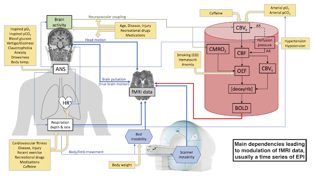
There are three broad categories of fluctuations or biases imprinted in the fMRI data. I've tried to depict them in Figure 1. At top-right, in a cartoon red blood vessel, is the cascade of physiological events leading to BOLD contrast. Next, on the left, there are perturbations arising from the subject's body. Some of these are direct effects, like head motion, and some are propagated via modulation of the same physiological parameters that give rise to BOLD. Breathing is a good example of the latter. A change in breathing depth or frequency can change the arterial concentration of CO2, leading to non-neural BOLD changes. Furthermore, the breathing rate is intricately tied to the heart rate, via the vagus nerve, and so
we can also expect altered brain pulsation. In the final category, depicted in my figure as scanner-based mechanisms at the bottom, we have experimental imperfections. In the last group are things that could be reduced or eliminated in principle, such as thermal drift in the gradients, wobbly patient beds, and resonance frequency shifts across the head arising from changing magnetic susceptibility of the chest during breathing. The thin blue lines connecting the different parts of the figure are supposed to show the main influences, with arrowheads to illustrate the directionality.
 |
| (Click image to enlarge.)
Figure 1.
Major routes of modulation in time series data in an fMRI experiment.
The flow chart in the depiction of a blood vessel, in red, is based on a
figure from Krainik et al. 2013
and shows the main events leading to BOLD via neurovascular coupling.
Main body-based mechanisms originate on the left, and scanner-based
experimental imperfections are depicted on the bottom. All mechanisms
ultimately feed into the fMRI data, depicted at center. Yellow boxes
contain some of the main modulators of mechanisms that can produce
either fluctuations or systematic biases in fMRI data.
Abbreviations: ANS - autonomic nervous system, HR - heart rate, CBVa - arterial cerebral blood volume, CBVv - venous cerebral blood volume, CMRO2 - cerebral metabolic rate of oxygen utilization, CBF - cerebral blood flow, OEF - oxygen extraction fraction, deoxyHb - deoxyhemoglobin, AR - autoregulation, pO2 - partial pressure of oxygen (O2 tension), pCO2 - partial pressure of carbon dioxide (CO2 tension). |
As if that wasn't already a lot of complexity, I'm afraid there's more. In the yellow boxes of Figure 1 are some of the main modulators of the underlying mechanisms responsible for perturbing fMRI data. These modulators are usually considered to be confounds to the main experimental objective. I posted a list of them a few years ago. Caffeine is probably the best known. It modulates both the arterial cerebral blood volume (CBVa) as well as the heart rate (HR). We already saw that HR and breathing are coupled, so this produces a third possible mechanism for caffeine to affect fMRI data. There's also an obvious missing mechanism: its neural effects. Some direct neural modulators are summarized in Figure 2, placed in their own figure simply to make this a tractable project. I'll be going back to reconsider any direct neural effects at the end of the series, to make sure I've not skipped anything useful, but my main emphasis is the contents of Figure 1.
 |
| Figure 2. Potential modulators of neural activity during an fMRI experiment. |
Measuring the modulators
There are about a dozen mechanisms leading to fluctuations in fMRI data. Note that some paths depicted in Figure 1 may contain multiple discrete mechanisms. The figure would be far too cluttered if every mechanism was depicted. Take head motion. It could be foam compressing through no fault of the subject, or it could be the subject fidgeting, or apparent head motion arising from the sensitivity of the EPI acquisition to off-resonance effects (for which there are at least two main contributions: thermal drift in the scanner and chest motion in the subject). I tried to estimate how many combinations are represented in Figure 1 but quickly gave up. It's several dozen. I'm not sure that knowing the number helps us. Clearly, it's an omelette.
So, what can we do about it? Well, there are only so many things one can measure before, during or after an MRI scan, so we should probably start there. In the first set of posts in this series I'll look at non-MRI measures that can be performed during fMRI data acquisition, to track moment to moment changes in some of the parameters of Figure 1. These will include:
- Heart rate
- Blood pressure
- Vascular low frequency oscillations in the periphery
- Respiration rate
- Expired CO2
- Electrodermal activity
- Eye tracking
- Head motion
Then, in the next set of posts I'll shift to assessing ancillary MRI measurements that can inform an fMRI experiment, such as:
- Anatomical scans
- Baseline CBF
- Blood oxygenation
- Cerebrovascular reactivity
- Calibrated fMRI (which is actually a slightly different way of doing the fMRI experiment, but requires some ancillary steps)
Finally, I'll consider informative, non-MRI data you could capture from questionnaires or relatively simple non-invasive testing. With better understanding, I am hoping that more researchers begin to consider physiology as earnestly as they do the domains involving psychology and statistics.